Retinal Vein Occlusions Specialist
Reading and Windsor, Berkshire
Expert RVO Treatment
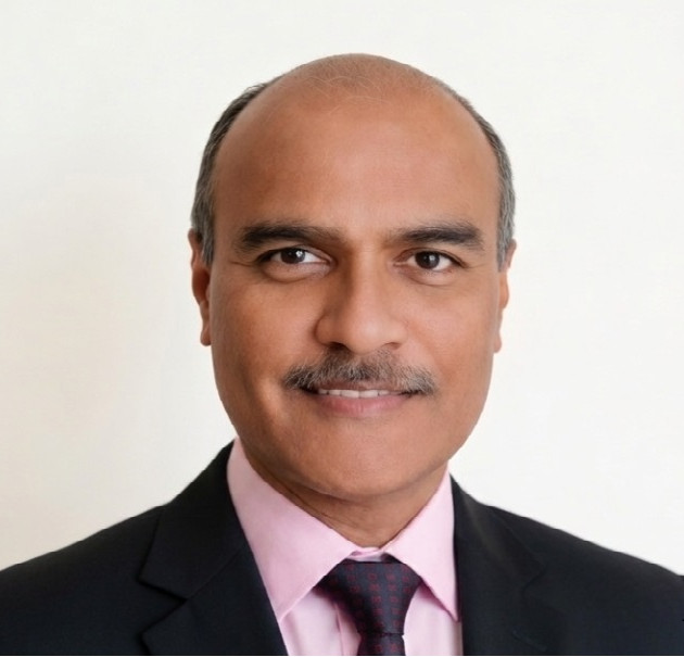
Mr Praveen Kumar is a senior NHS Consultant Ophthalmologist and Medical Retina Specialist providing private retinal vein occlusion (RVO) care in Berkshire and surrounding areas. With over 35 years of ophthalmology experience and extensive expertise in retinal disease, he offers specialist diagnosis, OCT imaging, retinal injections and laser treatment for retinal vein occlusion.
Retinal Vein Occlusion Care in Reading, Windsor and Berkshire:
Patients from Reading, Windsor, Maidenhead, Slough, Bracknell, Wokingham and surrounding areas can access specialist retinal assessment and treatment with Mr Kumar. His clinics provide consultant-led care for retinal vein occlusion, including urgent assessment for sudden changes in vision.
Understanding a Blockage in the Eye’s Blood Supply
The retina contains two main types of blood vessels: arteries, which supply oxygen-rich blood, and veins, which drain blood away from the eye. A healthy blood supply is essential for maintaining normal retinal function and clear vision.
Retinal vein occlusion (RVO), also known as a blocked retinal vein, occurs when one of the veins draining blood from the retina becomes blocked. The retina is the light-sensitive layer at the back of the eye responsible for vision. When blood flow is interrupted, fluid and blood can accumulate, causing retinal swelling and damage.
Without appropriate treatment, RVO can lead to reduced vision or, in some cases, permanent visual loss.
Types of Retinal Vein Occlusion (RVO)
Retinal vein blockage can occur at different levels of the retinal circulation:
-
Central Retinal Vein Occlusion (CRVO): A blockage of the main vein draining the retina.
-
Branch Retinal Vein Occlusion (BRVO): A blockage affecting one of the smaller retinal vein branches.
-
Hemiretinal Vein Occlusion: A blockage involving approximately half of the retinal circulation.
The impact on vision depends on the location and severity of the blockage, and whether the macula (the central part of the retina responsible for detailed vision) is affected.

Symptoms of Retinal Vein Occlusion
Symptoms vary depending on the severity and location of the blockage. Common symptoms include:
-
Sudden or gradual blurring of vision
-
Distorted vision, where straight lines may appear bent or wavy
-
A dark or missing area in the field of vision
-
Reduced sharpness of vision
-
Occasionally, significant sudden loss of sight
When to seek urgent help?
Sudden blurred vision, distortion, loss of vision, or a new dark area in your sight requires prompt retinal assessment. Early diagnosis allows appropriate treatment to be started quickly and may help reduce complications.
Any sudden change in vision should be assessed urgently by an eye specialist.
Retinal vein occlusion is often associated with conditions affecting blood vessels and circulation, including:
-
High blood pressure
-
Diabetes
-
Raised cholesterol
-
Age-related vascular changes
-
Glaucoma or raised eye pressure
-
Conditions affecting blood clotting or vascular health
Identifying underlying risk factors helps guide treatment and reduce future complications. In younger patients, further investigation may sometimes be required to identify less common causes.
-
Mr Kumar uses advanced retinal imaging and examination techniques to assess the severity of RVO and plan appropriate treatment.
Dilated retinal examination:
A detailed examination identifies retinal haemorrhages, swelling, areas of reduced blood supply and early signs of complications such as abnormal blood vessel growth.
OCT scan:
Optical coherence tomography (OCT) provides high-resolution images of the retinal layers, helping detect and monitor macular oedema, fluid leakage and response to treatment.
This non-invasive scan evaluates retinal blood flow, areas of ischaemia and vascular changes, helping assess disease severity and prognosis.
Fundus Fluorescein Angiography (FFA):
FFA uses an injected dye and retinal photography to assess blood flow, leakage, retinal damage and whether treatments such as injections or laser may be required.
With advanced retinal imaging and specialist medical retina expertise, Mr Kumar provides accurate diagnosis and evidence-based management of retinal vein occlusion.
Treatment depends on the type of occlusion, the effect on vision, and whether complications are present.
Management may include:
-
Eye injections to reduce retinal swelling and improve vision
-
Treatment to control abnormal blood vessel growth and prevent complications
-
Monitoring and treatment of raised eye pressure or associated glaucoma
-
Management of underlying vascular risk factors
Early assessment and appropriate treatment can help reduce complications and optimise visual outcomes.
-
Anti-VEGF Injections
Anti-VEGF injections are the most commonly used treatment for retinal vein occlusion. They work by blocking vascular endothelial growth factor (VEGF), a substance that increases when the retina is affected by poor oxygen supply and contributes to retinal swelling and fluid leakage.
Relying on his experience and results achieved, Mr Kumar routinely uses:-
Aflibercept 2mg (Eylea®)
-
Ranibizumab (Lucentis®)
-
Faricimab (Vabysmo®)
-
Eylea HD 8mg
-
Bevacizumab (Avastin®), in selected cases
Treatment usually involves an initial series of injections, followed by a personalised schedule based on retinal response and scan findings.
Steroid Implants
Steroid treatment may be considered in selected patients, particularly when swelling persists or response to anti-VEGF treatment is incomplete. Steroids reduce inflammation and leakage from retinal blood vessels.
• Dexamethasone implant (Ozurdex): A slow-release implant providing treatment over several months.
-
Retinal injections are performed as an outpatient procedure using local anaesthetic drops. The eye is cleaned carefully to minimise infection risk, and the medication is injected into the vitreous cavity.
Most patients tolerate the procedure well and return home shortly afterwards.
Regular follow-up with retinal examination and OCT imaging helps monitor treatment response and determine future management.
A typical visit includes:
-
Assessment of vision and symptoms
-
OCT retinal imaging
-
Review of scan results and treatment discussion
-
Intravitreal injection, if required
-
Planning of follow-up appointments
The injection itself takes only a few seconds, while the complete appointment usually lasts around 30–60 minutes.
-
Most patients experience only mild temporary symptoms following treatment. These may include:
-
Mild irritation or a gritty sensation
-
Watering of the eye
-
Slight redness at the injection site
-
Small floaters or tiny air bubbles that disappear naturally over the next day or two
Mr Kumar has performed thousands of retinal injections and provides advice and aftercare guidance to help minimise discomfort and identify any concerns early. Normal daily activities can usually be resumed the following day, although swimming is best avoided for several days after treatment.
Written aftercare instructions and emergency contact details are provided following each injection.
-
Without treatment, retinal swelling caused by RVO may persist and affect the macula, the area responsible for sharp central vision. Persistent swelling can result in worsening vision and, in some cases, permanent visual loss.
Severe cases with reduced retinal blood supply may also lead to abnormal fragile blood vessels developing inside the eye. These vessels can bleed or increase eye pressure, causing further complications.
Mr Kumar assesses the severity of each case and discusses the potential benefits and risks of treatment based on individual circumstances.
Delaying treatment may allow retinal swelling and fluid leakage to continue, increasing the risk of retinal damage and lasting visual impairment.
Early management can help control swelling and reduce complications related to poor retinal blood supply, including abnormal blood vessel growth.
Lasers
Pan Retinal Laser (PRP)
Panretinal photocoagulation (PRP) is a retinal laser treatment used in selected cases of RVO with significant retinal ischaemia (poor blood supply). It reduces the risk of complications by treating areas of the retina that may contribute to the development of abnormal fragile blood vessels.
PRP does not directly treat macular swelling but can help protect high-risk eyes from complications. Mr Kumar has extensive experience in retinal laser treatment and provides PRP when clinically appropriate.
Macular Grid Laser
Macular laser may be considered in selected cases of branch retinal vein occlusion (BRVO) with persistent macular swelling, particularly when other treatments are unsuitable or when fluid is away from the centre of the macula. Although anti-VEGF injections are now the main treatment for RVO-related macular oedema, laser remains useful in carefully selected patients.
Observation
Observation may be appropriate when vision is stable, there is no significant macular swelling, or immediate treatment is not required.
Regular retinal assessment and imaging help detect changes such as worsening swelling, reduced blood supply, or abnormal vessel growth. Treatment can then be started if the condition changes.Choosing an experienced retinal specialist is important for conditions that can affect vision. Mr Kumar provides consultant-led care for retinal vein occlusion, macular conditions, and other complex retinal disorders.
With extensive ophthalmology and medical retina experience, he offers advanced retinal imaging, evidence-based treatments, and individualised management plans. He receives referrals from NHS and private eye specialists for assessment, second opinions, and ongoing retinal care.
Through clinics in Reading and Windsor, patients across Berkshire and surrounding areas can access specialist retinal assessment and treatment focused on maintaining the best possible visual outcome.
Anti-VEGF therapy has significantly improved outcomes for patients with retinal vein occlusion by reducing swelling and helping many patients achieve better vision.
Mr Kumar uses modern licensed therapies guided by retinal scans, visual function, and treatment response.
Where appropriate, a Treat-and-Extend approach may be used to maintain disease control while reducing unnecessary visits. Treatment intervals are adjusted according to each patient’s needs, as some individuals require closer monitoring and more frequent treatment.
Retinal vein occlusion management continues to evolve with advances in imaging, research, and treatment.
Mr Kumar provides care based on current clinical evidence and international best practice. He applies knowledge from important retinal studies BRAVO, CRUISE, COPERNICUS, GALILEO, VIBRANT, and SCORE. Through ongoing engagement with retinal research, audits, and specialist conferences, he ensures patients benefit from modern, evidence-led treatment strategies.
He continues to engage with research, audits, and specialist education to ensure patients benefit from modern treatment approaches.
A key advantage of choosing Mr Kumar is continuity of care across both NHS and private practice.
As Lead Consultant for Macular Degeneration at King Edward VII Hospital, Windsor, and Senior Medical Retina Specialist at Royal Berkshire Hospital, Reading, he has extensive experience managing complex retinal conditions in both settings.
Patients may choose private assessment, continue NHS care, or transfer between pathways while remaining under the care of the same retinal specialist.
For chronic conditions such as retinal vein occlusion, continuity is valuable because treatment decisions depend on both current retinal findings and long-term disease patterns.
Mr Kumar is recognised and accredited by all major private medical insurance providers in the United Kingdom.
Patients with private medical insurance are encouraged to check their level of cover with their insurer prior to their appointment. His team can assist with queries regarding insurance authorisation, self-funding options, or transfer between private and NHS pathways.
Yes, it is usually possible to move between NHS and private eye care, depending on your individual circumstances and treatment needs.
If you are currently receiving NHS care and wish to have private treatment with Mr Kumar, you can arrange a private consultation. Your ongoing management can then be discussed and planned accordingly.
If you are receiving private care and wish to return to NHS services, this can also be arranged where appropriate.
Although NHS and private care can work alongside each other, they are separate pathways. Any change in care should be discussed carefully with your eye specialist to ensure continuity.